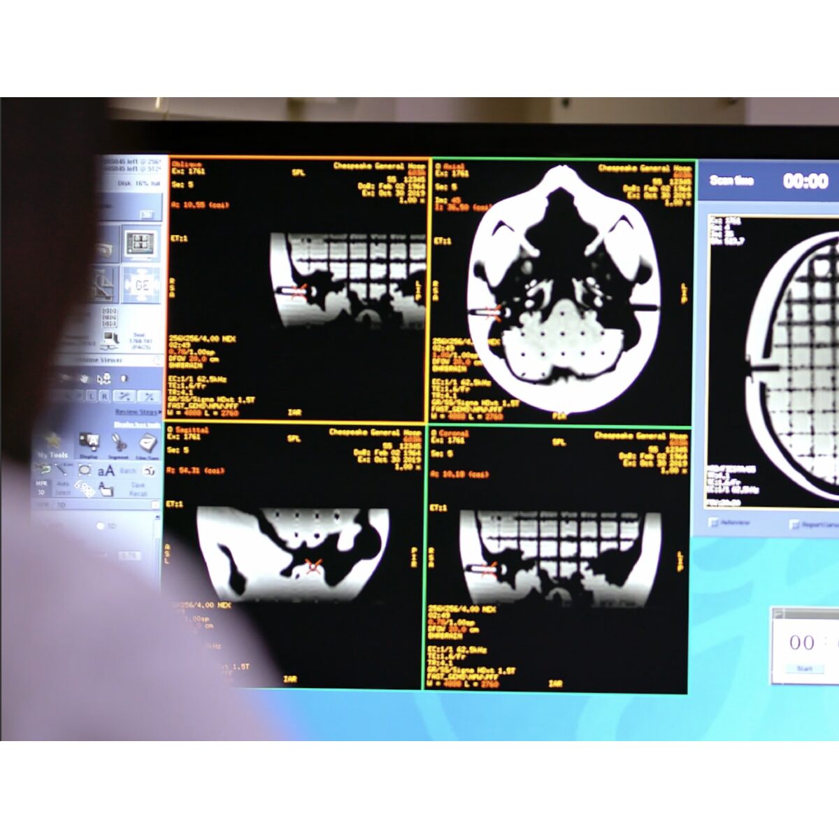

Characterize Geometric Accuracy for MR Use in Treatment Planning: The MR SRS Distortion Phantom is useful for verifying image fusion algorithms and deformable image registration used in various treatment planning systems. The anthropomorphic and tissue-equivalent design closely matches clinical imaging scenarios.
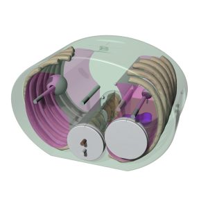
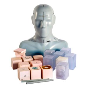
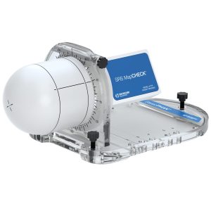
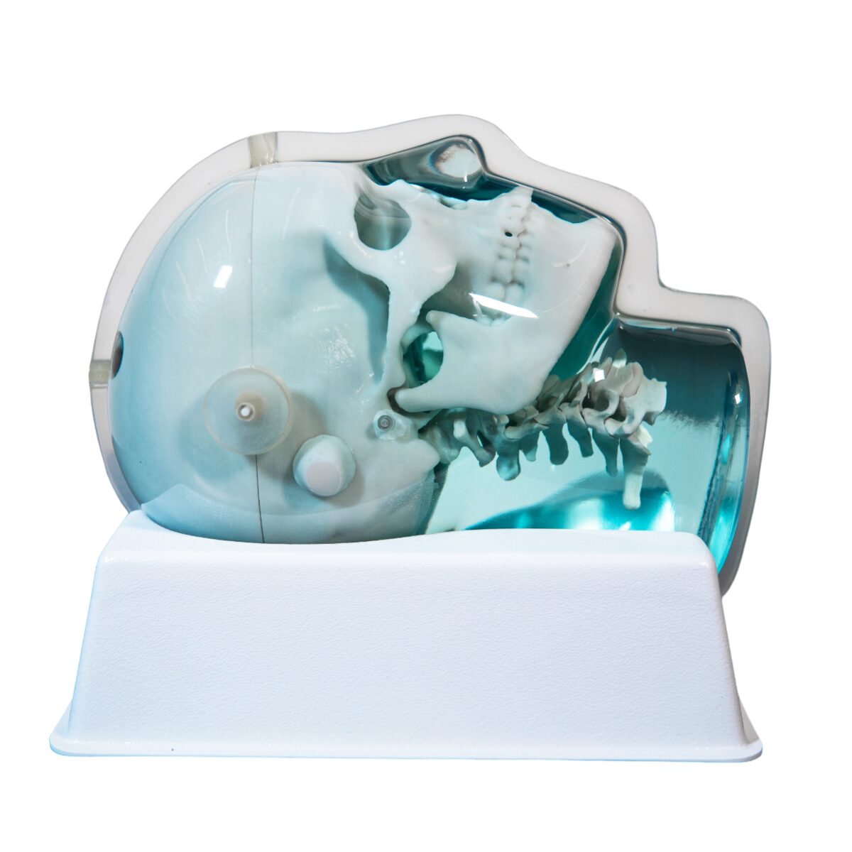
Anthropomorphic Tissue-Equivalent Head: The skull is made of a plastic-based trabecular bone substitute, and the interstitial and surrounding soft tissues are made of a proprietary water-based signal-generating polymer. The entire phantom is encased in a transparent plastic shell to protect the gel against desiccation. Specially designed pads allow fixation with any stereotactic frame or mounting devices for end-to-end testing. The phantom is also suitable for frameless stereotactic QA.
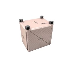
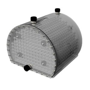
Detailed 3D Intracranial Design: The entire intracranial portion of the skull volume is filled with a 3D orthogonal grid of cross-shaped rods with a diameter of 2.5 mm spaced at 10 mm (I-S), 10.5 mm (AP), and 11 mm (L-R). Extra material added at grid intersections increases the grid signal. Five extended axis rods intersect at the grid reference origin. The end of each extended axis is equipped with CT/MR markers allowing precise positioning with lasers and co-registration of CT and MR image sets. The phantom contains air voids on both sides that replicate ear canals. These voids are used to evaluate common distortions encountered in clinical environments.
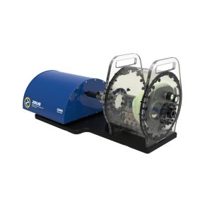
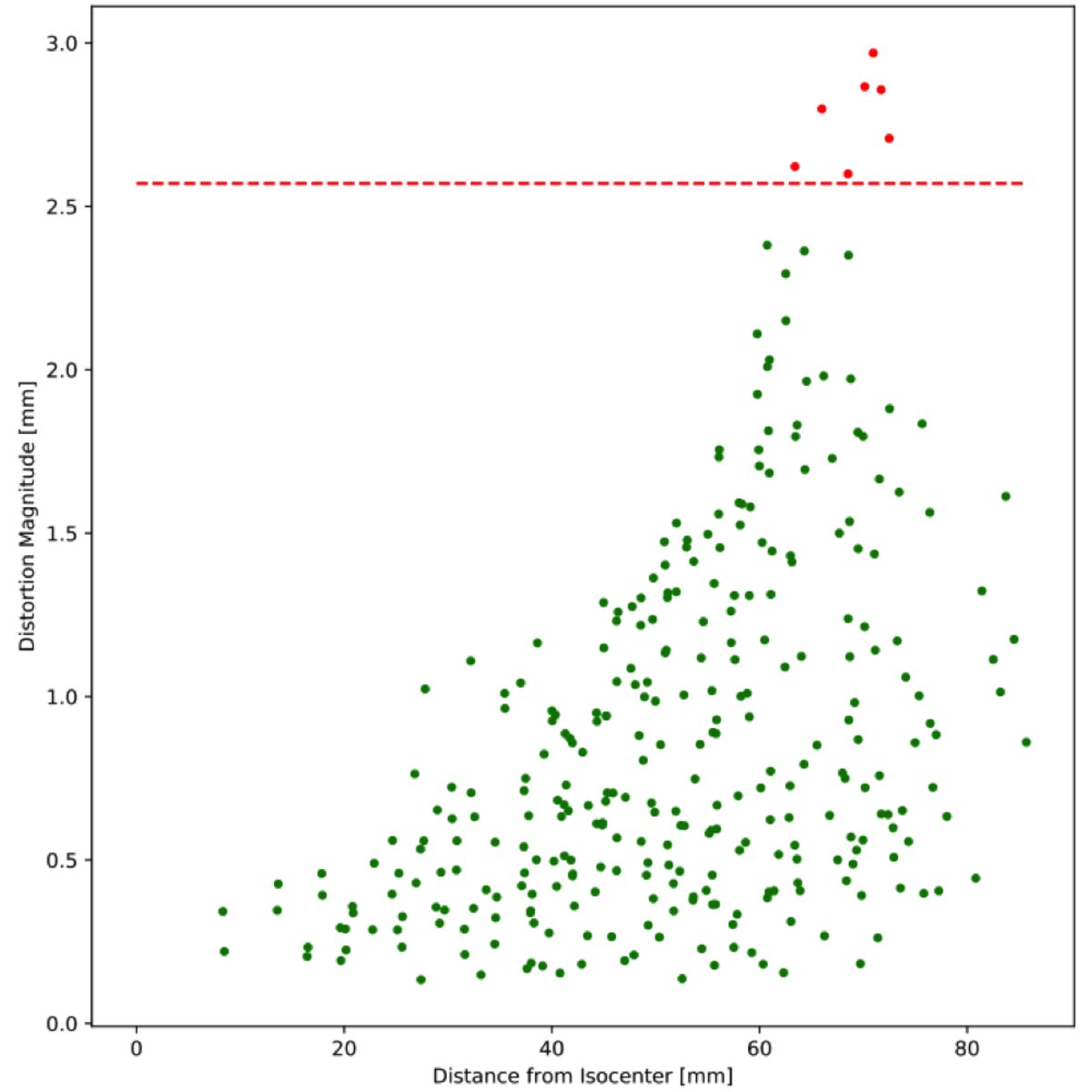
Automated Distortion Analysis in MRgRT: Used with MR grid phantoms, the Distortion Check software quickly and automatically quantifies distortion in MR images. Simply scan the phantom, load images, review reports and trend analyses, and export DICOM overlays. The software registers a ground truth CAD or CT scan to the detected control points. Then, interpolation is performed to generate the 3D distortion vector fields. Results can be reported in a variety of output formats, including scatter plots, contour plots, box and whisker plots for trend analysis, and DICOM overlays that can be exported to third-party software.
Software Features: Quickly and automatically analyze complete MR datasets Control point density optimized to approximate interpolation to linearity Easy-to-use cloud-based solution Detailed formatting in NEMA MS 12 standard recommendations Easily analyze and track multiple machines, image sequences, and phantoms Establish specific distortion tolerance limits for different image sequences.